«What began as a casual conversation between Marco Leona, David H. Koch Scientist in Charge of the Met's Department of Scientific Research, and Carol Stringari, Deputy Director and Chief Conservator of the Guggenheim Foundation, has grown over the past ten months into an unprecedented collaboration aiming to advance the role of science within curatorial and conservation-based scholarship at both institutions. The partnership—described in detail on the Guggenheim's blog—has established a framework for scientific research within the Guggenheim conservation studio by creating a position for the first scientist on staff and granting access to the Met's fully equipped chemical laboratories and advanced analytical instrumentation. Conservators and scientists from the two museums are currently sharing resources, identifying projects of mutual interest, and jointly studying objects in their respective collections.»
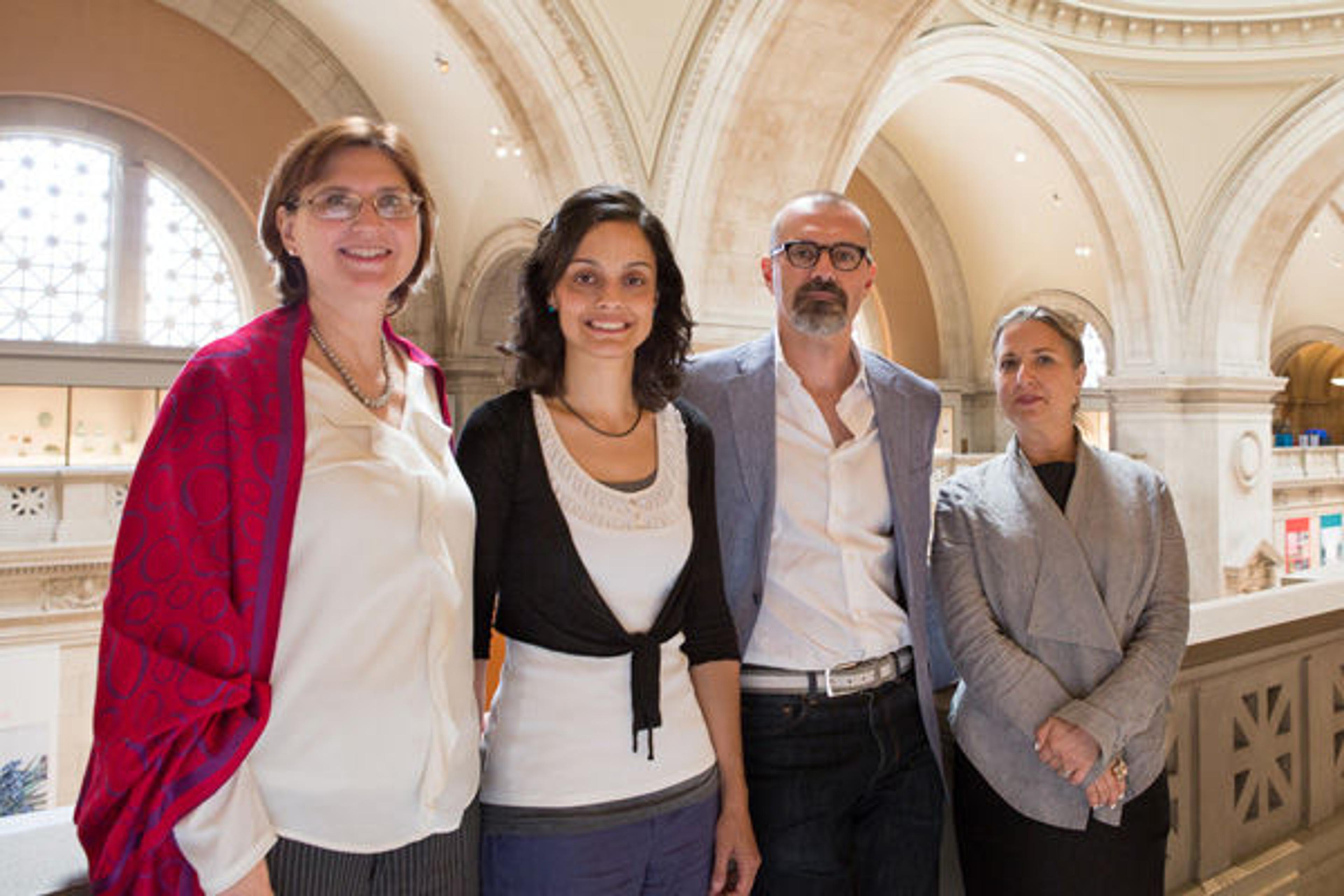
The collaborative team at the Met. Left to right: Julie Arslanoglu, Federica Pozzi, Marco Leona, and Carol Stringari
One of our first research endeavors as a collaborative team has been a comprehensive investigation of paintings by the Italian artist Alberto Burri (1915–1995) in support of the Guggenheim's upcoming exhibition Alberto Burri: The Trauma of Painting. We began this project by taking a joint look at a selection of Burri's works, defining several areas of inquiry, and designing a research plan that would address both art-historical and condition-related questions. We then inspected the paintings using both macroscopic and microscopic methods, as well as a wide range of analytical techniques; some of these techniques were noninvasive, meaning that they did not involve sampling the object under study, while some others required the removal of a microscopic specimen—typically the size of a grain of sand or smaller.
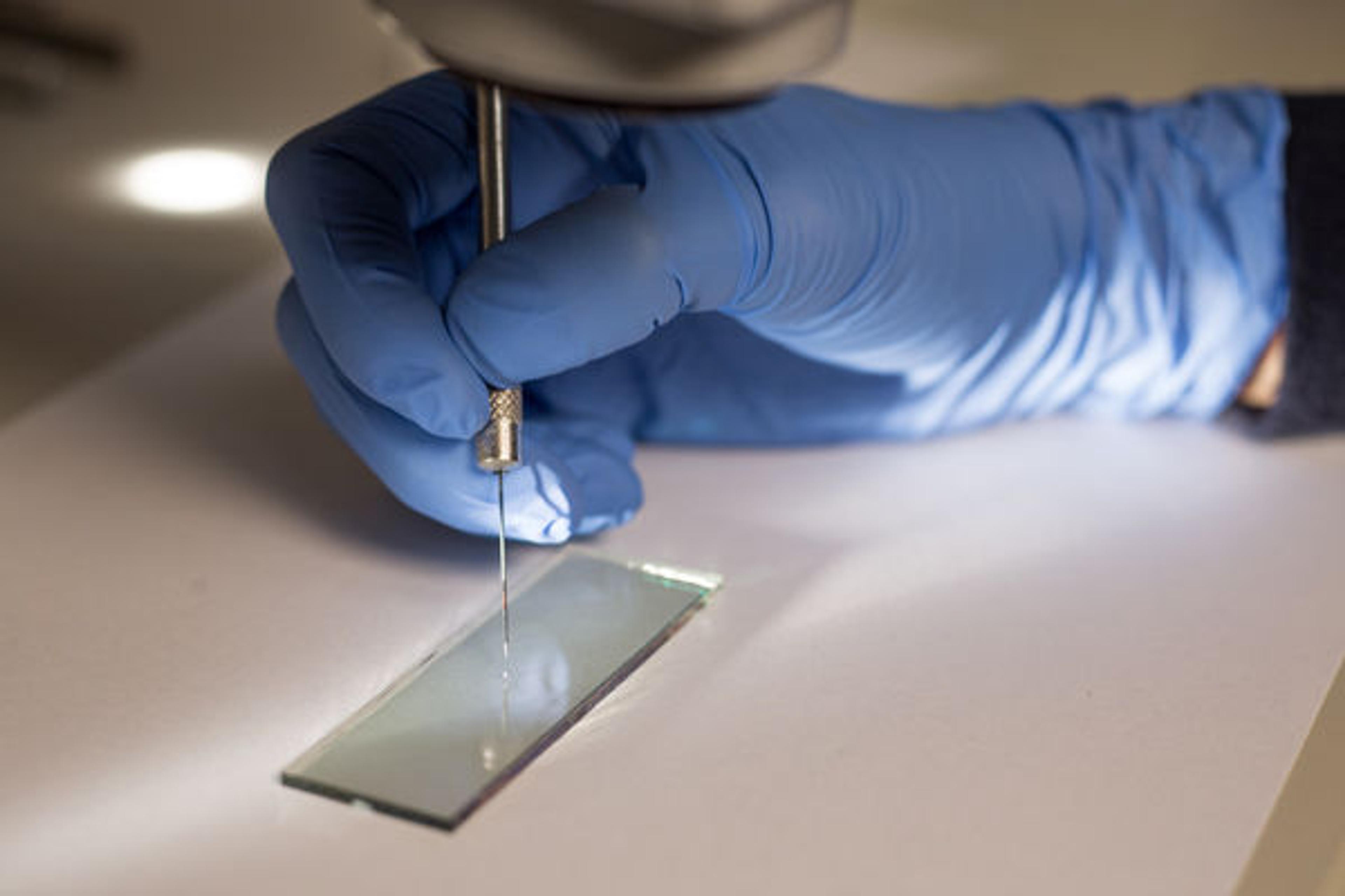
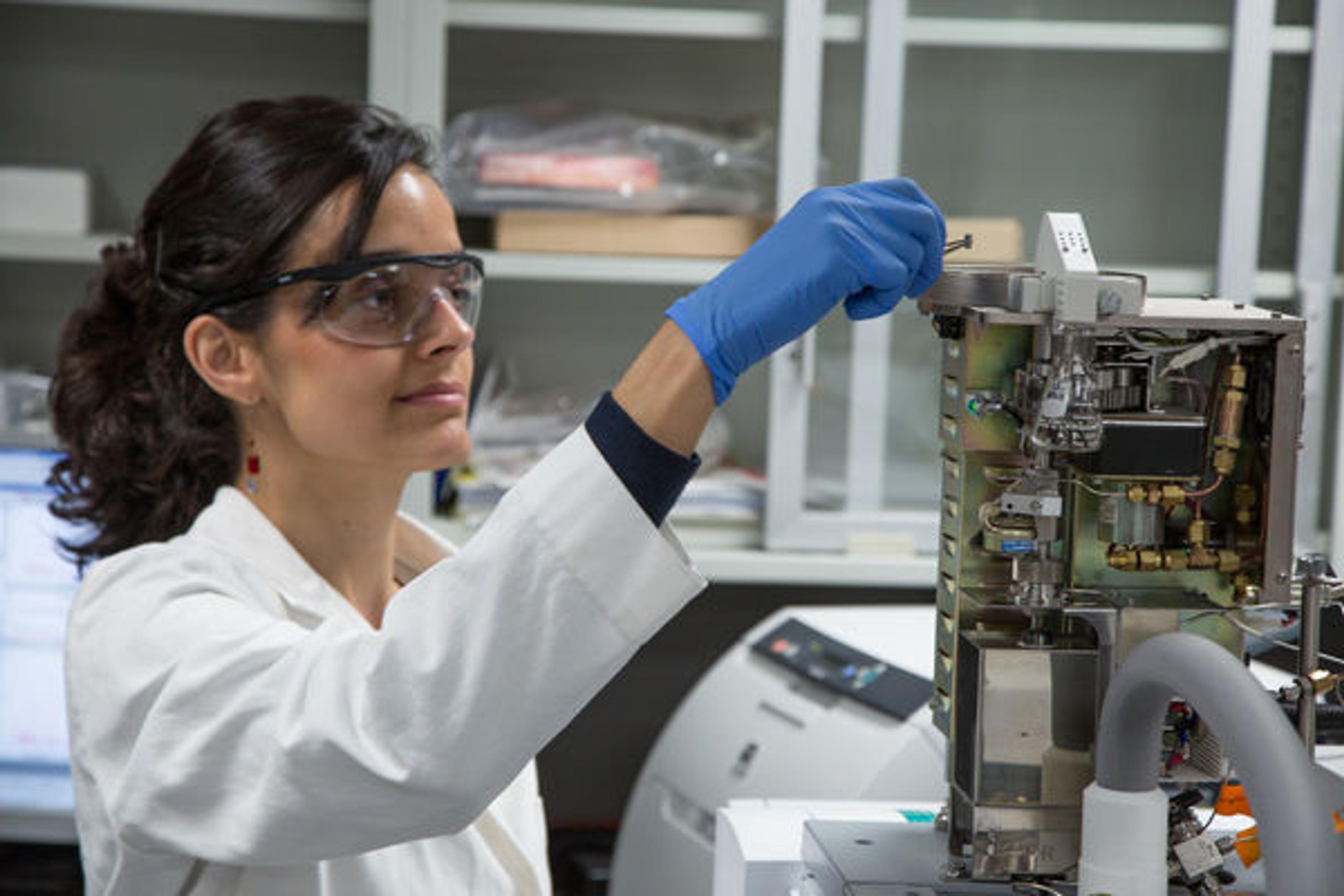
A minute sample of white paint, barely visible to the naked eye, is transferred onto a glass microscope slide for analysis with the aid of a tungsten needle.
In-depth scientific analysis of Burri's works was crucial to demonstrate how extensively the artist experimented with materials. He was profoundly influenced by the introduction to the market of new synthetic polymers, which he incorporated, along with traditional pigments and other common industrial products, into his creations. In some cases, the examination of cross sections—microscopic samples mounted in a resin block and polished to show the painting's stratigraphy—has revealed the presence of several paint layers under the artwork's surface, indicating extensive reworking and gradual changes in the composition's formal design. The information provided by the different analytical techniques we used was often complementary and, once interpreted in its entirety, enabled us to account for the incredible complexity of color shades, textures, surface appearances, and other physical features displayed by the artworks examined.
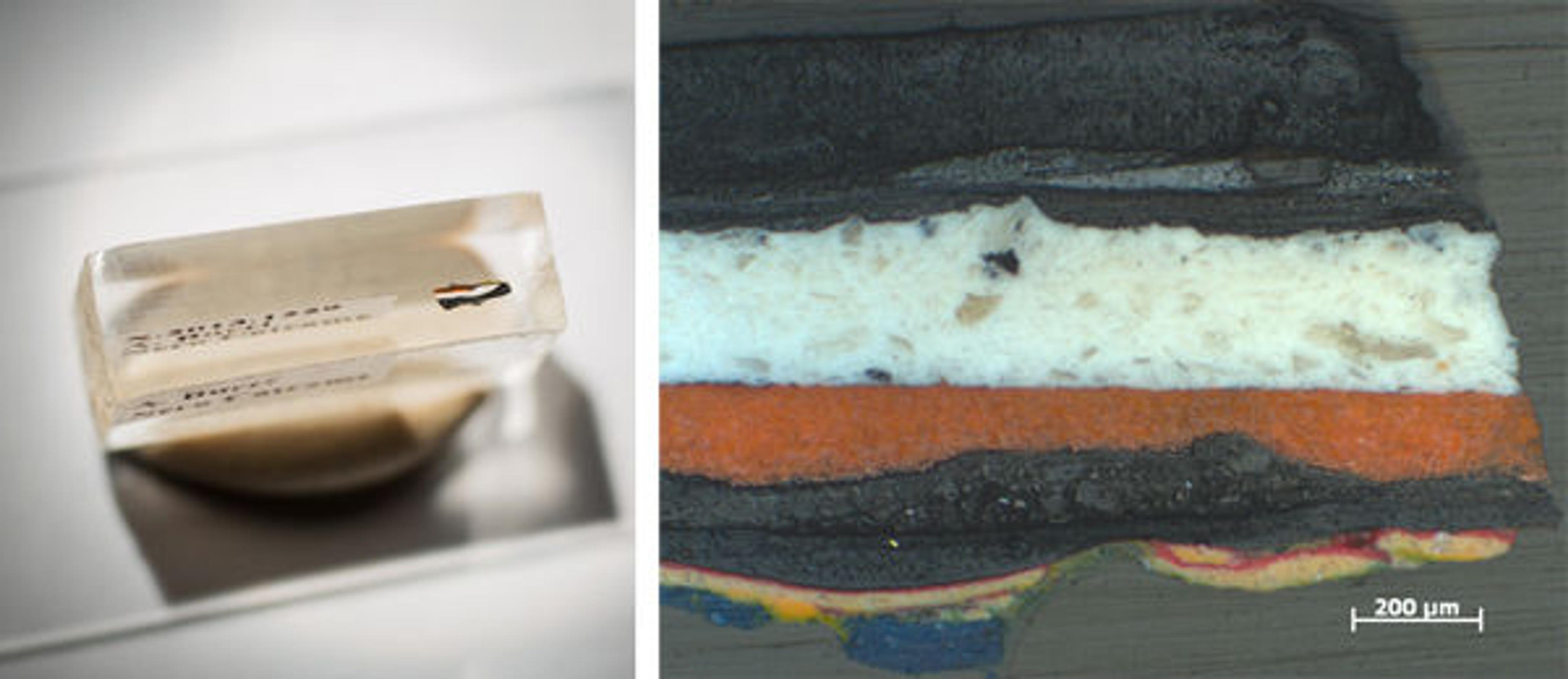
Left: A paint sample from Burri's Nero Catrame (1950) is embedded in methyl methacrylate resin and mounted as a cross section for materials analysis. Right: Polarized light image of the sample (100x magnification), showing several brightly colored layers below the thick bituminous surface.
Burri's Muffa T (1952), for instance, includes a variety of white paints ranging from extremely cold hues to much warmer cream tones, each incorporated in smooth, lumpy, dry, crumbly, cracked, matte, glossy, opaque, or reflective surfaces. Initial noninvasive analysis through X-ray fluorescence (XRF) spectroscopy revealed that all these white paints are zinc-based, with the occasional addition of other elements in minor or trace amounts. We were able to confirm the presence of zinc white, a common nineteenth- and twentieth-century artist's pigment, using Raman spectroscopy to examine minute samples taken from the painting. We also discovered—using Raman and Fourier-transform infrared (FTIR) spectroscopy—the presence of barite, calcite, gypsum, and titanium white; it appears that Burri was combining these materials with zinc white in various proportions to achieve particular color tonalities, surface textures, and translucency effects.

Left: Alberto Burri (Italian, 1915–1995). Muffa T (Mold T), 1952. Oil, PVA, pumice, sand, and shellac on canvas; 90.2 x 110.5 cm. Godwin-Ternbach Museum, Queens College, City University of New York (CUNY), Gift of G. David Thompson, 1958. Right: Micrograph of a small region of the painting at far right, showing cracked-white, smooth-black, and resinous taffy-colored paints above a mold-like area.
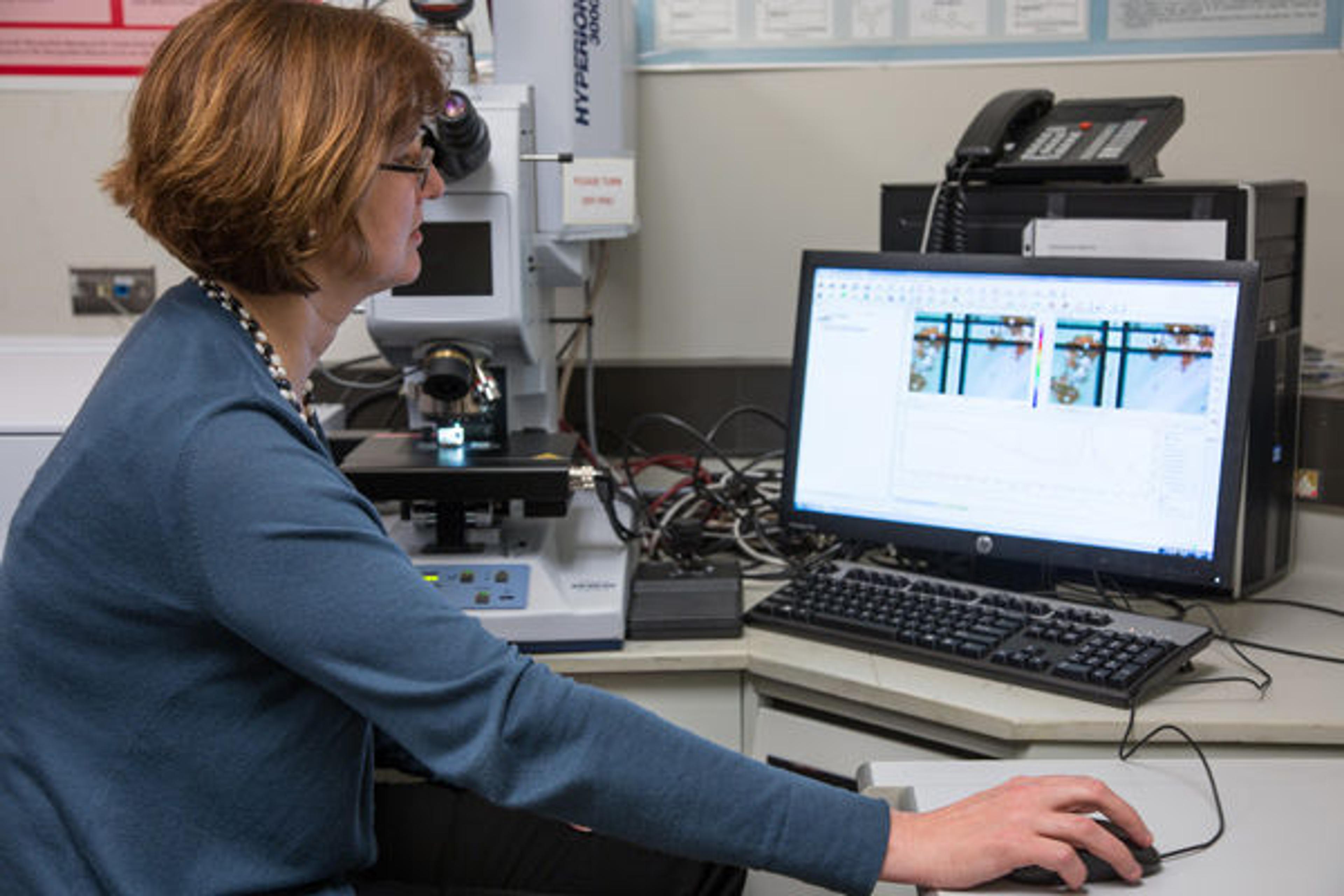
Arslanoglu analyzes the paint materials in a cross section sample using attenuated total reflection (ATR)-FTIR spectroscopy.
Another analytical method known as gas chromatography/mass spectrometry (GC/MS), which is especially suitable for the identification of binding media—the chemical components that bind pigments together to form paint—showed that Burri selectively used newly manufactured materials such as polyvinyl acetate (PVA), along with modified oils, either individually or in combination, to lend particular visual and tactile properties to different areas of the artwork. The relative amounts of pigments and binding media also play a key role in determining the final appearance of the paint surface and texture in certain regions. This is particularly evident for Muffa T, where highly reflective, medium-rich areas contrast with fissures that result from overly dry paint mixtures with high pigment/binder ratios and anticipate the much later Cretti series.
Pozzi loads a paint specimen into the gas chromatography/mass spectrometry (GC/MS) autosampler for binding media analysis.
Another fascinating example of Burri's liberal experimentation is the mold-like accretions that permeate the surface of Muffa T with ingenious sculptural effects. In a curious blend of associations, some of these areas evoke the dampness and humidity of Burri's native Umbria, while the granular, sandy, desiccated consistency of adjacent areas recalls the arid desert landscapes familiar to the artist from his time spent in East and North Africa and the Texas panhandle.
From a materials perspective, the idea of mold growing on the painting surface is rendered through sludgy accumulations of dense pictorial matter. Our analysis revealed that this "mold efflorescence" was built up using composite conglomerations of pumice stone, calcite, gypsum, quartz and other silicate minerals, mixed in with an abundance of brass flakes, which we identified by scanning electron microscopy/energy-dispersive X-ray (SEM/EDS) analysis. Interestingly, the detection of atacamite, a copper(II) chloride hydroxide, in green parts of these areas suggested that such green coloration might have been achieved through the oxidation of the brass flakes rather than by the addition of specific green pigments.
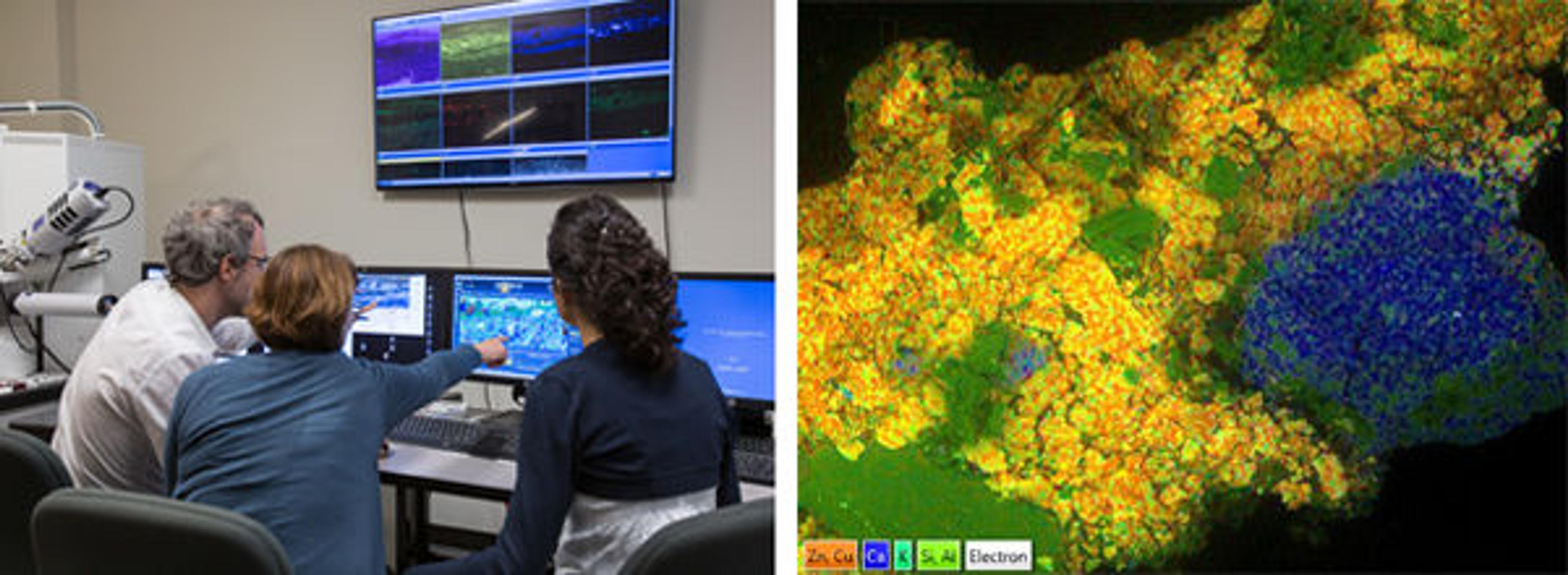
Left: Arslanoglu and Pozzi, together with Met scientist Federico Carò, examine paint cross sections at the scanning electron microscope (SEM). Right: Composite X-ray map of a portion of a paint sample from Burri's Muffa T (1952), where the distribution of copper and zinc (brass flakes), calcium (calcite), silicon, aluminum, and potassium (pumice fragments and other silicate grains) is highlighted.
Ultimately, our scientific analysis of Burri's paintings has provided a deeper understanding of the multifaceted nature of the artist's working practice. Having examined several of his works, we found consistent material choices and a recurring visual lexicon, both of which originate from extensive experimentation and a skillful amalgamation of various components. More detailed results of this technical study will be included in the exhibition catalogue, which, hopefully, will serve as a guide for the viewer to appreciate the works from a different perspective. This highly complex and fascinating exhibition will grace the walls of the Guggenheim rotunda from October 9, 2015, to January 6, 2016. Don't miss it!
Related Link
Guggenheim Blogs: "Art and Science on Fifth Avenue: The Met and the Guggenheim Combine Forces" (July 15, 2015)